


5 days ago2 min read


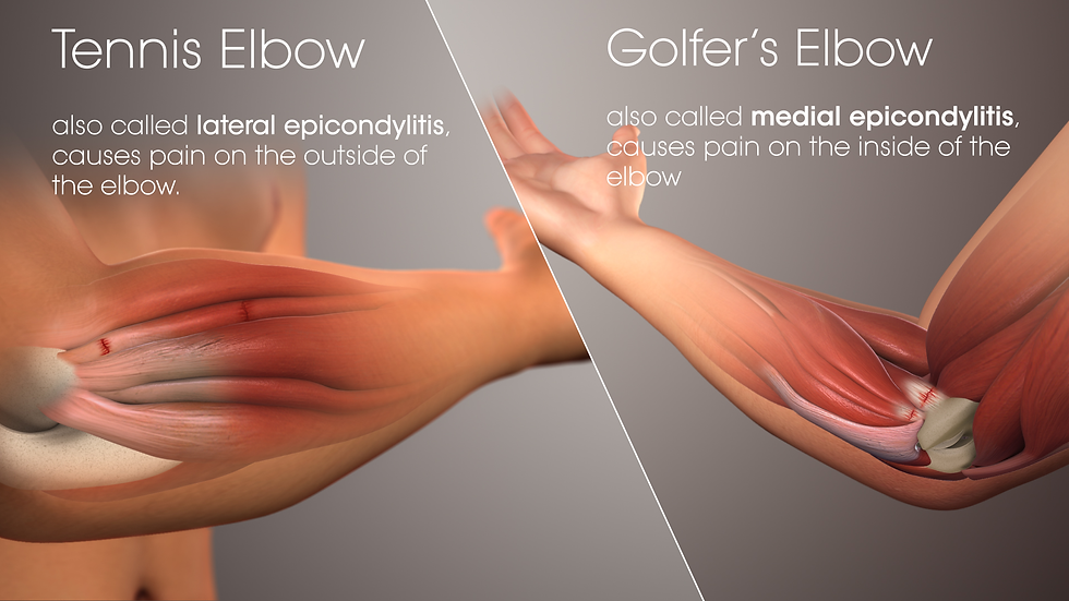
Lateral Epicondylitis or TENNIS ELBOW
→ Described by Runge in 1873
It is a self-limiting degenerative tenodesis of the common extensor origin of the elbow.
→ ECRB is the most common involved.
(as it it relatively tortious throughout the origin.
Anatomy
Lateral epicondyle gives origin to the extensor group of muscles.
The extensors are order of BR-ECRL- ECRB - EDL-ECU
the ECRL origin is muscular from the origin itself
ECRB is tendonous for long distance
PATHOGENESIS
Stresses over a tendon
👇🏻
Tendon tolerates the stress by stretching
👇🏻
Cross linkage and collagen deposition.
👇🏻
Reduced stretching
👇🏻
when stress continues and it crosses its
👇🏻
tolerance limit.
👇🏻
microtear
👇🏻
Continuous stress leads to multiple tears.
👇🏻
Tendinosis
👇🏻
Angiofibroblastic hyperplasia (a manifestation
of granulation tissue)
👇🏻
This disturbs normal collagen synthesis
👇🏻
Necrosis and fibre regeneration.
extension of wrist extensor
* pain with resisted wrist extension
* pain with gripping activities
* decreased grip strength
* palpation & inspection
- [ ] point tenderness at ECRB insertion into lateral epicondyle few mm distal to tip of lateral epicondyle
- [ ] neuromuscular
* May have decreased grip strength
* Neurological exam helps to differentiate from entrapment syndromes
* Radiograph (recommended views)
* AP/Lateral of elbow
Usually normal, may reveal calcifications in the extensor muscle mass (up to 20% of patients) may reveal signs of previous surgery
* MRI
not necessary for diagnosis
increased signal intensity at ECRB tendon origin may be seen (up to 50% of cases)
Ultrasonography
* Requires experienced operator (variable sensitivity/specificity)
* Most useful diagnostic tool in experienced operator hands
* ECRB tendon appears thickened and hypoechoic
1. Rest
2. Physiotherapy -stretching and strengthening (better than rest and reduced activity if done adequately
* Increase strength of the shoulder and scapula for better elbow function
* Strengthening of scapular stabilisers-lower trapezius, serratus anterior and rotator cuff muscle
3. Epicondylar counterforce braces - reduce tension over extensors. Can cause secondary nerve problems due to prolonged use
4. Non-steroidal anti-inflammatory drugs (NSAIDs)
5. Corticosteroid injections
* Control local inflammatory response and pain medication.
* Useful in initial phases- 4 weeks
* Potential side-effects including changes in colouration of the skin, fat atrophy and muscle wasting.
6. Autologous blood injections
* Stimulate inflammatory response and bring in the necessary nutrients to promote healing.
* Useful in recalcitrant cases.
7. Platelet-rich plasma injections (PRP).
* contain high concentrations of growth factors,
* enhance tendon healing.
8. Percutaneous radiofrequency thermal treatment.
* inducemicrotenotomy and remove pathological tissue.
9. Extracorporeal shock-wave therapy
10.Acupuncture
11. Botulinum toxin A injections
Remember most of the above mentioned modalities have only short term results
Operative treatment
Indications -
* persistent pain and disability despite well-performed conservative treatment
Principle
* debriding the angio-fibrotic tissue of the ECRB with or without posterior tendon repair.


Comments